Osteoporosis
Osteoporosis is a systemic disease characterized by bone mass loss, a violation of the bone tissue microarchitectonics and is accompanied by increased bone fragility and an increased risk of fractures. When the bone and bone mass is formed, bone density of the skeleton becomes stable at the age of 20. Starting from the age of 35, it gradually decreases. Age atrophy constitutes about 1% per year.
Bone tissue loss particularly worsens after menopause, fluctuating within 3 to 5% per year. By the age of 75, the bone tissue loss constitutes about 50% in the spine and 20 to 30% in the tubular bones.

The main bone strength determinant is bone mass. According to bone mineral density (BMD) measurements, lumbar spine osteoporosis prevails in people who are 50 years of age and older at 19.8% ratio in women and 13.3% in men; femoral neck osteoporosis – 21.9% in women and 21.7% in men. Densitometry reveals signs of osteoporosis at the age of 50 to 59 in 2.4% of the examined women, at the age of 60 to 69 – in 10.3%, and over the age of 70 – in 14.7%. Bone mass loss accelerates in people after the age of 60.
Osteoporosis is dangerous for its complications – fractures occurring not only as a result of a trauma, often a minor one, but also spontaneously. Though the fracture union occurs, it remains insufficient, especially in the spongy bone where new bone growth is not possible to occur in a proper way since the bone does not contain epiphysial surfaces in the form of the periosteal coverage which the compact bone contains. The risk of repeated fractures caused by even minor traumas significantly increases. This also happens due to the ligamentous apparatus strength decrease and lower resistance to loads in intervertebral discs.
Both the trabecular heads and the cylindrical diaphyseal cortical bone section, which form the long bones, are also affected by osteoporosis.
In Russia, 14 million people (10% of the population) suffer from osteoporosis, and fractures during life develop in 6% of the population. A significant part of these fractures is caused by the bone strength decrease due to osteoporosis. Fractures lead to long-term disability and life quality deterioration. Femoral neck fractures, common in older people, in 40% of cases lead to a fatal outcome within a 6- month period. 90% of these femoral neck fractures in elderly people are osteoporotic in nature. The world population ageing leads to further increase in the incidence of osteoporosis. Preventive measures, early diagnosis, and treatment of osteoporosis will allow millions of people to prolong their active life.
Osteoporosis risk factors are classified into modifiable (potentially changeable) and non-modifiable.
Modifiable factors:
- vitamin D deficiency;
- alcohol abuse;
- tobacco smoking;
- low physical activity;
- low body mass index and/or low weight;
- low calcium consumption;
- predisposition to falling.
Non-modifiable factors:
- age over 65;
- hypogonadism in men and women;
- long-term immobilization;
- Caucasoid race;
- female sex;
- low mineral tissue density;
- previous fractures;
- glucocorticoids intake;
- early (including surgical) menopause in women.
Besides, a number of diseases, as well as the intake of medications affecting bone tissue metabolism are additional risk factors for the secondary osteoporosis development.
Clinical picture
The disease develops gradually, and it is asymptomatic for long periods. The first manifestations of osteoporosis are fractures that occur with minor traumas (e.g., falling from one’s own body height) or spontaneously.
For osteoporosis, the dorsal and lumbar vertebrae, the distal forearm bones, and the proximal femur fractures are most typical.
In vertebral fractures, a decrease in bone growth is noted when compared to growth at a young age (from 1 to 3 cm with a single vertebra fracture, from 9 to 15 cm with multiple fractures). Vertebral fractures are accompanied by acute (with compression fracture) or chronic (with gradual vertebral bodies settling caused by own weight) pain in the back, thoracic hyperkyphosis and lumbar lordosis worsening. The waistline gradually disappears and the abdomen is protruded forward, and in severe cases, the lower ribs touch the iliac wings.
Pain syndrome is accompanied by severe functional impairments limiting the daily activity of the patient.
Bone strength measurement
The bone strength or bone resistance to fractures is determined by bone mineral density, microstructure, elasticity, bone surface size, and cortical layer thickness.
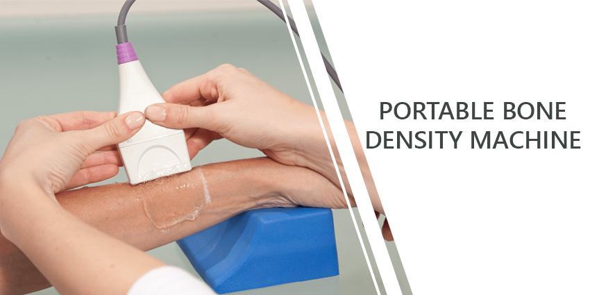
There are two main methods to diagnose osteoporosis: dual-energy X-ray absorptiometry (DXA) which is used to determine bone mineral density (BMD), and bone ultrasound examination which measures the speed of ultrasound through the bone (SOS).
Both methods (DXA and ultrasound) allow determining the bone tissue strength and the risk of fractures with sufficient accuracy. According to the International Society for Clinical Densitometry (ISCD) recommendations, ultrasound examinations are indicated for a wide population segment in different osteoporosis risk groups.
Based on the examination results, a number of patients can be appointed to undergo examinations using the X-ray DXA densitometer (Quantitative Ultrasound in the Management of Osteoporosis: The 2007 ISCD Official Positions).
Osteoporotic vertebral fractures diagnosis should be based on a combination of symptoms, including complaints, anamnesis, objective examination data, and risk factor assessment, and be carried out using radiographic methods.
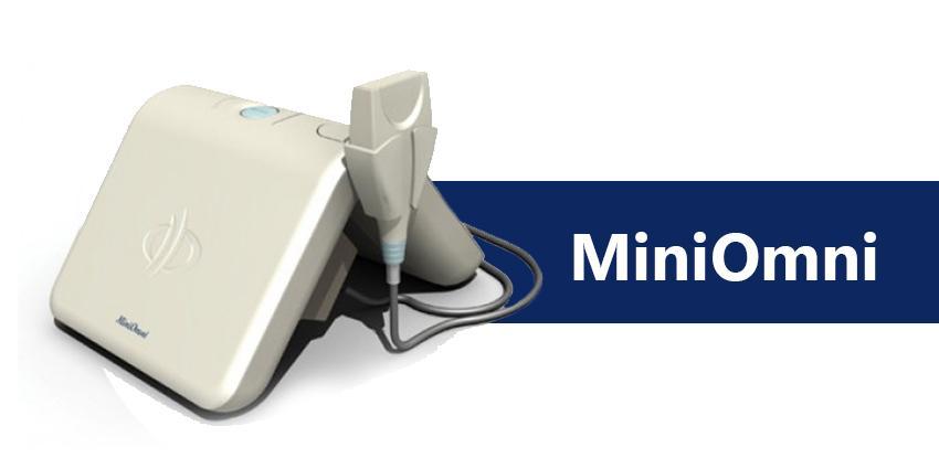
Due to the zero ionizing radiation, ultrasound devices, for example, the Israeli ultrasound densitometer MiniOmni, allow examining children and women across pregnancy, as well as to repeatedly carry out examinations to monitor the treatment.
MiniOmni devices, unlike other ultrasound densitometers, examine not the heel bone, but those bones which are the most susceptible to fractures (tibia, ray, phalanx, and metatarsal). Besides, MiniOmni allows carrying out densitometry to newly-born children.
All these features bring the capabilities of the MiniOmni ultrasound densitometer to the capabilities of a much more complex and expensive dual-energy X-ray densitometers (DXA).
Bone strength tissue measurement based on WHO criteria
The bone strength measurement, using any method, is carried out according to two types of parameters:
Ø T score – The difference between the patient’s SOS index and the average SOS index in the population of young healthy people, measured in units which are equal to the standard deviation from the mean peak value.
Ø Z score - The difference between the patient’s SOS index and the average SOS index in a population of the same age and sex as the patient, measured in units equal to the standard deviation from the mean peak value.
Based on the disease prevalence, the World Health Organization (WHO) has set the criteria used to determine the risk of fractures and to diagnose osteoporosis. According to these criteria, the osteoporosis diagnosis is made based on the patient’s T score:
|
Normal healthy bones Osteopenia Osteoporosis Severe osteoporosis |
T score is more than -1,0 T score is in the range from -1.0 to -2.5 T score is below -2.5 T score is below -2.5 and there is evidence of fractures with minor traumas |
Treatment
General recommendations for all patients include:
- balanced calcium and vitamin D intake;
- lifestyle changes aimed at physical exercises strengthening the muscles and reducing body weight;
- smoking cessation; alcohol dependence detection and treatment, as well as the treatment of other fracture risk factors, for instance, visual impairment.
Sources:
- Rheumatology. National guide / eds V.A. Nasonova, E.L. Nasonov. – M.: GEOTAR-Media, 2008, – 720 p.
- Osteoporosis. Clinical recommendations. 2nd ed. / eds O.M. Lesnyak, L.I. Benevolenskaya. – M.: GEOTAR-Media, 2010, – 272 p.
- National Osteoporosis Foundation. Clinician’s Guide to Prevention and Treatment of Osteoporosis. – Washington, DC: National Osteoporosis Foundation, 2010. – 44 p.
- WHO publication: Kanis J.A., on behalf of the World Health Organisation Scientific Group. Assessment of osteoporosis at the primary health care level. WHO Collaborating. Centre for Metabolic Bone Diseases. – University of Sheffield, 2007. – 338 p.
- Rachner T.D., Khosla S., Hofbauer L.C. Osteoporosis: now and the future // Lancet. – 2011. – Vol. 377 (9773). – Р. 1276–1287.
05.03.2018